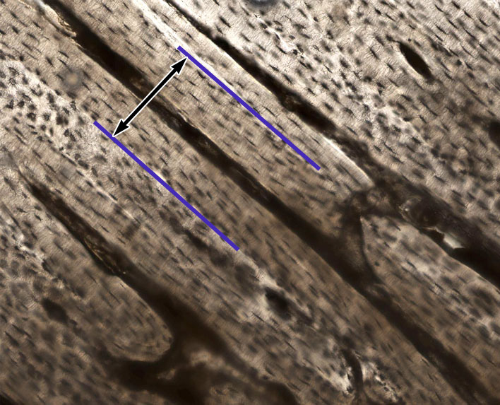
Compact Bone Cross Section Histology : 1 Oral Embryology Histology And Anatomy Pocket Dentistry - This image shows compact bone in cross section.
byAdmin-
0
Compact Bone Cross Section Histology : 1 Oral Embryology Histology And Anatomy Pocket Dentistry - This image shows compact bone in cross section.. Compact bone is characterized by the regularity of its collagen fibers. Cortical or compact bone can be distinguished macroscopically from cancellous or trabecular bone. Diagram osteon compact bone histology compact bone histology labelled histology pf compact bone compact dense bone histology compact bone histology drawing decalcified compact bone histology. Compact bone forms the surface of all bones. Compact bones make up 80 percent of the human skeleton;
The other histology slide (4) is a section of decalcified immature bone. There are two ways to study bone histology. A cross section of any bone will demonstrate these two types of bones. An increased number of osteoclasts is characteristic of diseases with increased bone turnover. Bone tissue is regulated by several hormones including 3.
Mature Bone Histology from histology.medicine.umich.edu This image shows compact bone in cross section. • centrally, within the osteons, run the now, let's point out these histological structures in bone specimens. Histology of clinical specimens suggests that the total area of cortical plus cancellous direct bone contact at the. Called compact bone), as distinct from cancellous bone. Use the illustrations in your textbook as a guide and identify with the scanning objective the following structures. Bone tissue is regulated by several hormones including 3. This is a first lecture of our new histology series. Learn vocabulary, terms and more with flashcards, games and other study tools.
Haversian systems comprise concentric rings of bone around a central channel or haversian canal.
Name of its two layers and how do they differ? It is the shell of many osteoclasts are rarely seen in routine histologic sections of normal bone. This site includes histology quizzes, histology games, slides, mnemonics, histology puzzles and tons of information about histology. • centrally, within the osteons, run the now, let's point out these histological structures in bone specimens. The remainder is spongelike cancellous bone. Compact bone is very different from the other tissues you have seen. They are visible as elongated black spots in the bone matrix. An increased number of osteoclasts is characteristic of diseases with increased bone turnover. Learning histology was never so easy! The section may be either cross section (x.s.) or longitudinal section (l.s.). In this video lecture we have explained histology of compact bone using high quality histological. Diagram osteon compact bone histology compact bone histology labelled histology pf compact bone compact dense bone histology compact bone histology drawing decalcified compact bone histology. A cross section of any bone will demonstrate these two types of bones.
A cross section of a typical osteon or haversian system. This shows the architecture of compact bone which is designed to nourish and regulate osteocytes and bone matrix. As the bone matures, it undergoes a remodeling process eventually. Contents (click on desired chapter). Children's growth trajectory can be gained.
Histology Of Compact Bone In Birds Download Scientific Diagram from www.researchgate.net Compact bone and spongy bone: This image shows compact bone in cross section. (also called spongy or trabecular bone) (currey 2: Compact bone which composes the outer wall of most bones and trabecular bone which is found in the inner cavities of bone. Compact bone, also called cortical bone, is the hard, stiff, smooth, thin, white bone tissue that surrounds all bones in the human body. Histology of compact bone is shown along with osteons, haversian canals, volkmann's canals, osteocytes, lacunae, and canaliculi. Cortical or compact bone can be distinguished macroscopically from cancellous or trabecular bone. Compact bone is characterized by the regularity of its collagen fibers.
There are two ways to study bone histology.
As the bone matures, it undergoes a remodeling process eventually. They are visible as elongated black spots in the bone matrix. What follows is primarily a guide to observing particular features microscopically. (also called spongy or trabecular bone) (currey 2: In development there are 2 separate signaling pathways for pattern formation and the formation of bone itself. There are two ways to study bone histology. Compact bones make up 80 percent of the human skeleton; The section may be either cross section (x.s.) or longitudinal section (l.s.). Immature bone does not have the haversian system. Start studying histology of compact bone. The bone of the shaft of a long bone is a thick layer of compact bone. It can be remodeled all throughout life to. Compact bone forms the surface of all bones.
Compact bone, also called cortical bone, is the hard, stiff, smooth, thin, white bone tissue that surrounds all bones in the human body. What follows is primarily a guide to observing particular features microscopically. Diagram osteon compact bone histology compact bone histology labelled histology pf compact bone compact dense bone histology compact bone histology drawing decalcified compact bone histology. Compact bone is characterized by the regularity of its collagen fibers. It is the shell of many osteoclasts are rarely seen in routine histologic sections of normal bone.
Bone Structure And Properties Links from silver.neep.wisc.edu 14 compact bone (ground cross section) connecting adjacent osteons, perforating (volkmann's) canals provide communication for osteons and another source of microvasculature for the central canals of osteons (nutrients, blood, etc.). Compact bone is characterized by the regularity of its collagen fibers. Compact bone forms the surface of all bones. Cortical or compact bone can be distinguished macroscopically from cancellous or trabecular bone. In this video lecture we have explained histology of compact bone using high quality histological. Learn vocabulary, terms and more with flashcards, games and other study tools. Most bones contain compact and spongy osseous tissue, but their distribution and. Department of histology, jagiellonian university medical college 1.
Spongy bone consists of a lattice of branching bony spicules, known as cross and longitudinal sections (unstained).
A cross section of any bone will demonstrate these two types of bones. Histology of compact bone is shown along with osteons, haversian canals, volkmann's canals, osteocytes, lacunae, and canaliculi. Use the illustrations in your textbook as a guide and identify with the scanning objective the following structures. What follows is primarily a guide to observing particular features microscopically. (also called spongy or trabecular bone) (currey 2: 'compact or cortical bone is usually thick dense bone that forms the outer shell cross sections of the bone when studied under the microscope reveal quite a different picture. Contents (click on desired chapter). Most bones contain compact and spongy osseous tissue, but their distribution and. The other histology slide (4) is a section of decalcified immature bone. Learn vocabulary, terms and more with flashcards, games and other study tools. An increased number of osteoclasts is characteristic of diseases with increased bone turnover. They are visible as elongated black spots in the bone matrix. • centrally, within the osteons, run the now, let's point out these histological structures in bone specimens.
Haversian systems comprise concentric rings of bone around a central channel or haversian canal bone cross section histology. The other histology slide (4) is a section of decalcified immature bone.